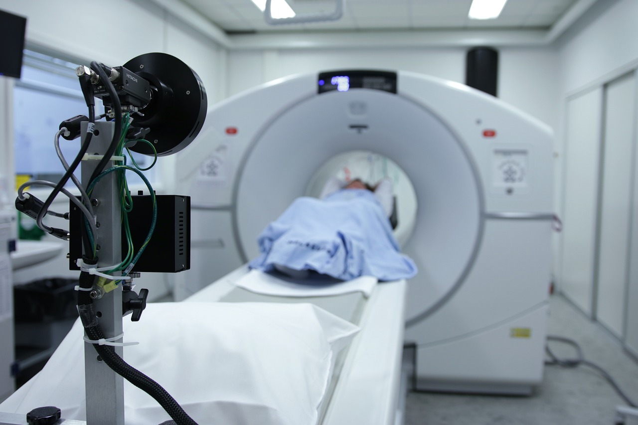
CT (computed tomography) scans are linked to an increased risk of blood cancers in children, a new study says.
According to the study, published in Nature Medicine, a single CT scan elevates the risk of blood cancer by 16%. One million people under the age of 22 were evaluated as part of the study.
“In terms of absolute risk, this means that, for every 10,000 children who have a CT scan, we can expect to see about 1-2 cases of cancer in the 12 years following the examination,” said Magda Bosch de Basea, lead researcher of the study.
CT scans are computerized X-ray imaging procedures used for the diagnosis of several diseases, including cancer. It is a lifesaving tool that helps in the easy identification of tumors, abnormalities and injuries in the body. During the procedure, the body gets exposed to ionizing radiation, which in low doses does not cause health risks.
Around five to nine million CT scans are performed every year on children in the U.S. The procedure accounts for the largest contributor to medical radiation exposure.
According to the National Cancer Institute, the risk of radiation from CT scans is more concerning for children than adults. The heightened risk is due to higher sensitivity to radiation and their longer life expectancy, giving them a larger timeframe for expressing radiation damage. Children are also at risk of receiving higher doses of radiation if settings are not adjusted for their body size.
For the study, researchers looked into data from nine European countries – Belgium, Denmark, France, Germany, Netherlands, Norway, Spain, Sweden and the U.K. The health of the participants was tracked for nearly eight years.
A single CT scan accounts for an average dose of eight milligrays, which is associated with bone marrow damage. The researchers found that multiple CT scans or accumulation of radiation to 100 milligrays triples the risk of developing blood cancer.
“The exposure associated with CT scans is considered low, but it is still higher than for other diagnostic procedures,” said Elisabeth Cardis, a study author. “The procedure must be properly justified – taking into account possible alternatives – and optimized to ensure that doses are kept as low as possible while maintaining good image quality for the diagnosis.”


